Wikiversity Journal of Medicine/Tubal pregnancy with embryo
< Wikiversity Journal of Medicine| |
This work is peer reviewed. The peer review statement is located at . A permanent link to the peer-reviewed version is located at . |
Author: Ed Uthman
PDF version (checked)
|
Suggested citation format:
First submitted: 20 July 2014 Accepted: 20 July 2014 Last updated: 29 February 2016 Licensing:
|
| |
Media in this article is on display in Wikipedia in the following articles: Atavism, [https://en.wikipedia.org/wiki/Ectopic pregnancy Ectopic pregnancy], Embryo, Human, [https://en.wikipedia.org/wiki/Prenatal development Prenatal development] |
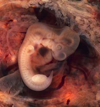
Human embryo (7th week of pregnancy, 5th week p.o.)
This photo of an opened oviduct with an ectopic pregnancy features a spectacularly well preserved 10-millimeter embryo. It is uncommon to see any embryo at all in an ectopic, and for one to be this well preserved (and undisturbed by the prosector's knife) is quite unusual.
Even an embryo this tiny shows very distinct anatomic features, including tail, limb buds, heart (which actually protrudes from the chest), eye cups, cornea/lens, brain, and prominent segmentation into somites. The gestational sac is surrounded by myriad chorionic villi resembling elongated party balloons. The age of this embryo is 7 weeks of gestational age.
The photo was taken on Kodak Elite 200 slide film, with a Minolta X-370 camera and 100mm f/4 Rokkor bellows lens at near-full extension. The formalin-fixed specimen was immersed in tap-water and pinned to a tray lined with black velvet. The exposure was 1/4 second at f/8.