Wikiversity Journal of Medicine/Ultrasonography of a cervical pregnancy
< Wikiversity Journal of Medicine| |
This work is peer reviewed. The peer review statement is located at . A permanent link to the peer-reviewed version is located at . |
Author: Jesper Agrell
|
Suggested citation format:
First submitted: 15 November 2014 Accepted: 15 November 2014 Last updated: 29 February 2016 Licensing:
|
Author correspondence: jesper.agrell![]() lvn.se
lvn.se
PDF version (checked)
| |
Media in this article is on display in Wikipedia in the following article: [https://en.wikipedia.org/wiki/Cervical pregnancy Cervical pregnancy] |
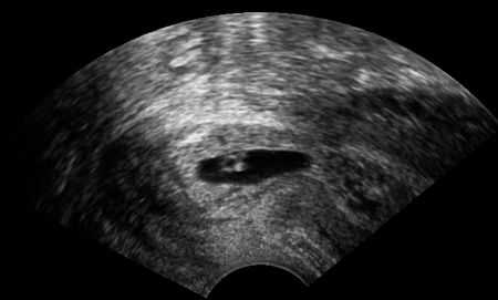
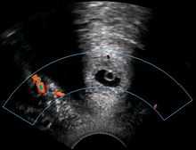
A woman in her 20s came to the gynecologic clinic because of a positive pregnancy test and a history of a previous ectopic pregnancy. The vaginal ultrasonography performed by Dr. Jesper Agrell showed a gestational sac in the cervix as displayed in the sagittal plane. The corpus of the uterus is located at right in the image. There was a discernible heartbeat, and the gestational sac diameter corresponded to a gestational age of 5 weeks. The distance from the gestational sac to the external orifice was only 15 millimeters.