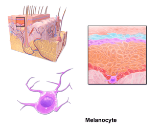
This computer drawing models a dermal melanocyte and its dendrites. Credit:
BruceBlaus.
Melanocytes are ink cells, from the Greek μέλανo-ink, or black ink and χυτός-cast, for cell.
Genetics
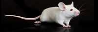
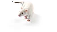
This is a white, null mouse that has been knocked-out for tyrosinase. Credit: The Jackson Laboratory.
In humans, a melanogenic enzyme, tyrosinase, is produced by the gene, GeneID: 7299. Actually, this gene produces a precursor to the enzyme. The precursor is activated once inside a closed melanosome.
"The enzyme [EC 1.14.18.1] encoded by this gene [GeneID: 7299] catalyzes the first 2 steps, and at least 1 subsequent step, in the conversion of tyrosine to melanin. The enzyme has both tyrosine hydroxylase and dopa oxidase catalytic activities, and requires copper for function."[1]
Theoretical melanocytes
A melanocyte is a cell containing the pigment melanin, which has several forms. A cell which yields or produces melanin, or one of its forms, may be called a melanoparagocyte, where parag- or parago- is from παραγ-yield or produce, or παραγο-producing.
Visuals
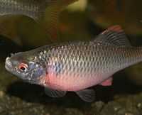
The dark appearance of the dorsal side of the male bitterling
Rhodeus amarus is caused by a dispersal of melanosomes in simulate the dark bottom of the fish tank. Credit:
Viridiflavus.
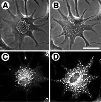
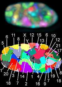
Def. a "cell containing melanin or other black pigment, such as are found in fish, amphibians, and reptiles"[2] is called a melanophore.
The image at the right shows a melanophore containing melanosomes (A), dispersed melanophores (B), fluorescent undispersed melanophores (C), and fluorescent dispersed melanophores (D).
The melanosomes have been dispersed out into the cells dendrites.
As an example, the image at the left shows a male bitterling Rhodeus amarus where the dark appearance of its dorsal surface is produced by a dispersal of melanosomes to simulate the dark bottom of the fish tank.
Hypotheses:
- Each location of a melanocyte has a genetically unique melanocyte.
- Melanoblasts can be signaled to divide with daughter cells migrating to locations formerly occupied by melaoncytes, e.g. dermal matrix.
This is an image of a Lewis rat. Credit: Charles River Laboratories.
The findings demonstrate a statistically systematic change from the status quo or the control group.
“In the design of experiments, treatments [or special properties or characteristics] are applied to [or observed in] experimental units in the treatment group(s).[3] In comparative experiments, members of the complementary group, the control group, receive either no treatment or a standard treatment.[4]"[5]
Def. a “short and/or incomplete realization of a certain method or idea to demonstrate its feasibility"[6] is called a proof of concept.
Def. evidence that demonstrates that a concept is possible is called proof of concept.
The proof-of-concept structure consists of
- background,
- procedures,
- findings, and
- interpretation.[7]
See also
References
- ↑ NCBI (12 June 2014). "TYR tyrosinase [ Homo sapiens (human) ]". Bethesda, Maryland USA: National Institutes of Health. Retrieved 2014-07-08.
- ↑ Visviva (27 November 2008). "melanophore, In: Wiktionary". San Francisco, California: Wikimedia Foundation, Inc. Retrieved 2014-07-08.
- ↑ Klaus Hinkelmann, Oscar Kempthorne (2008). Design and Analysis of Experiments, Volume I: Introduction to Experimental Design (2nd ed.). Wiley. ISBN 978-0-471-72756-9. http://books.google.com/?id=T3wWj2kVYZgC&printsec=frontcover.
- ↑ R. A. Bailey (2008). Design of comparative experiments. Cambridge University Press. ISBN 978-0-521-68357-9. http://www.cambridge.org/uk/catalogue/catalogue.asp?isbn=9780521683579.
- ↑ "Treatment and control groups, In: Wikipedia". San Francisco, California: Wikimedia Foundation, Inc. May 18, 2012. Retrieved 2012-05-31.
- ↑ "proof of concept, In: Wiktionary". San Francisco, California: Wikimedia Foundation, Inc. November 10, 2012. Retrieved 2013-01-13.
- ↑ Ginger Lehrman and Ian B Hogue, Sarah Palmer, Cheryl Jennings, Celsa A Spina, Ann Wiegand, Alan L Landay, Robert W Coombs, Douglas D Richman, John W Mellors, John M Coffin, Ronald J Bosch, David M Margolis (August 13, 2005). "Depletion of latent HIV-1 infection in vivo: a proof-of-concept study". Lancet 366 (9485): 549-55. doi:10.1016/S0140-6736(05)67098-5. http://www.ncbi.nlm.nih.gov/pmc/articles/PMC1894952/. Retrieved 2012-05-09.
External links
| Anthropology resources |
|---|
| | Activities | | | | Articles | | | | Categories |
Agriculture ·
Anthropology ·
Archaeology ·
Art ·
Arts ·
Biological Anthropology ·
Culture ·
Dominant group ·
Education ·
Evolutionary anthropology ·
Evolutionary psychology ·
Genetics ·
History ·
Humanities ·
Language ·
Linguistics ·
Literature ·
Medicine ·
Mythology ·
Philosophy ·
Psychology ·
Religious studies ·
Semantics ·
Social anthropology ·
Social psychology
| | | Courses | | | | Fields | | | | Glossaries | | | | Lectures | | | | Lessons | | | | Lists |
| | | Original research | | | | Portals |
History ·
Social sciences
| | | Problem sets | | | | Projects | | | | Proposals | | | | Quizzes | | | | Schools |
Agriculture ·
Alternative medicine ·
Anthropology ·
Archeology ·
Classics ·
Dentistry ·
Gastronomy ·
History ·
Life ·
Medicine ·
Pharmacy ·
Veterinary medicine
| | | Topics | |
|
| Gene project |
|---|
| | Activities |
| | | Articles | | | | Categories |
Agriculture ·
Biochemistry ·
Biology ·
Genetics ·
Medicine ·
Molecular evolution ·
Research projects ·
Zoology
| | | Courses | | | | Lectures | | | | Lessons | | | | Lists | | | | Original research | | | | Projects | | | | Proposals | | | | Quizzes | | | | Schools |
Agriculture ·
Alternative medicine ·
Biology ·
Brewing ·
Chemistry ·
Conservation Sciences ·
Dentistry ·
Gastronomy ·
Life ·
Marine sciences ·
Medicine ·
Pharmacy ·
Plant sciences ·
Veterinary medicine ·
Zoology
| | | Topics | |
|
| Humanities resources |
|---|
| | Activities | | | | Articles | | | | Categories |
Agriculture ·
Anthropology ·
Archaeology ·
Art ·
Arts ·
Culture ·
Dominant group ·
Education ·
Genetics ·
History ·
Humanities ·
Language ·
Linguistics ·
Literature ·
Medicine ·
Philosophy ·
Psychology ·
Religious studies ·
Semantics ·
Social psychology
| | | Courses | | | | Fields | | | | Glossaries | | | | Lectures | | | | Lessons |
| | | Lists |
| | | Original research | | | | Portals |
History ·
Social sciences
| | | Problem sets | | | | Projects | | | | Proposals | | | | Quizzes | | | | Schools |
Agriculture ·
Alternative medicine ·
Anthropology ·
Archeology ·
Classics ·
Dentistry ·
Gastronomy ·
History ·
Life ·
Medicine ·
Pharmacy ·
Veterinary medicine
| | | Topics | |
|
| Materials science resources |
|---|
| | Activities | | | | Articles | | | | Categories |
Astronomy ·
Astronomy Project ·
Astrophysics ·
Atmospheric science ·
History of science ·
Materials science ·
Metallurgy ·
Physics ·
Sciences ·
Technology
| | | Courses | | | | Glossaries |
| | | Lectures | | | | Lessons |
| | | Original research | | | | Problem sets | | | | Projects | | | | Quizzes | | | | Schools |
Chemistry ·
Construction ·
Engineering ·
Materials Science and Engineering
| | | Topics |
Amateur astronomy ·
Astronomy ·
Astrophysics ·
Electron microscopy ·
Geophysics ·
Materials science ·
Materials science and engineering ·
Metallurgical engineering ·
Nanoscience ·
Nanotechnology
Nuclear physics ·
Particle physics
|
|
| Medicine resources |
|---|
| | Activities | | | | Articles | | | | Categories |
Anatomy ·
Anesthesiology ·
Basic sciences ·
Blood tests ·
Cardiovascular medicine ·
Clinical Skills ·
Clinical trials ·
Complementary and alternative medicine ·
Dentistry ·
Dermatology ·
Diseases ·
Emergency Medicine ·
Endocrinology ·
Evidence-based Medicine ·
Gastroenterology ·
Geriatric medicine ·
Gerontology ·
Health ·
Healthcare ·
Hematology ·
Medical biophysics ·
Medical emergencies ·
Medical physics ·
Medical subject headings ·
Medicinal plants ·
Medicinal plants and wild foods ·
Medicine ·
Medicine stubs ·
Military Medicine ·
Nephrology ·
Neuroprotection ·
Oncology ·
Ophthalmology ·
Orthopedics and Rheumatology ·
Otolaryngology ·
Otorhinolaryngology ·
Pain ·
Pathology ·
Pharmacy ·
Physiology ·
Podiatry ·
Primary Care Medicine ·
Proctology ·
Psychiatry ·
Regenerative medicine ·
Reproductive medicine ·
Respiratory medicine ·
Risk management ·
School of Medicine Brainstorming ·
Urology
| | | Courses | | | | Glossaries | | | | Lectures | | | | Lessons | | | | Lists | | | | Problem sets | | | | Projects |
Gene Project ·
Medicine/Projects
| | | Quizzes | | | | Schools |
Medicine/Schools ·
Alternative medicine ·
Biology ·
Dentistry ·
Gastronomy ·
Medicine ·
Nursing ·
Pharmacy ·
School Health ·
Veterinary medicine ·
Zoology
| | | Syndromes | | | | Topics |
Medicine/Topics ·
Anesthesiology ·
Angiology ·
Anatomy ·
Basic sciences ·
Biology ·
Biomedical gerontology ·
Biomedicine ·
Cardiovascular medicine ·
Chemistry ·
Clinical skills ·
Community medicine ·
Cytogenetics ·
Emergency Medicine ·
Endocrinology ·
Epidemiology ·
Evidence-based Medicine ·
Gastroenterology ·
General surgery ·
Genetics ·
Geriatric Medicine ·
Health Informatics ·
Hematology ·
Histology ·
Immunology ·
Internal medicine ·
Laboratory Techniques ·
Medical physics ·
Medicinal botany ·
Medicinal chemistry ·
Military medicine ·
Nephrology ·
Neurology ·
Oncology ·
Ophthalmology ·
Orthopedics and Rheumatology ·
Otorhinolaryngology ·
Pathology ·
Pediatric Medicine ·
Pharmacology ·
Physiology ·
Primary Care Medicine ·
Psychiatry ·
Psychology ·
Regional Anatomy ·
Reproductive Medicine ·
Respiratory Medicine ·
Rheumatology ·
Serology ·
Virology ·
|
|
| Phosphate biochemistry |
|---|
| | Activities |
| | | Articles | | | | Courses | | | | History |
| | | Lectures |
Adenosine triphosphate ·
Bones ·
Cartilage ·
Cytoskeleton ·
Degenerate nucleotide ·
Deoxyribonucleic acid ·
Epigenome ·
Extracellular fluid ·
Ferredoxin ·
Flavodoxin ·
Human bones ·
Human teeth ·
Hydroxyapatite ·
Inorganic pyrophosphatase ·
Microfilaments ·
Microtubules ·
Nucleic acids ·
Nucleoside triphosphate ·
Organophosphate ·
Origin of life: polyphosphate ·
Oxidative phosphorylation ·
Phosphatases ·
Phosphate minerals ·
Phosphates ·
Phosphate biochemistry ·
Phosphate budget ·
Phosphate homeostasis ·
Phosphate reactions ·
Phosphate transistasis ·
Phospholipids ·
Phosphoramidates ·
Phosphoric acid ·
Phosphorites ·
Phosphorylation ·
Polyphosphates ·
Skeletons ·
Structural phosphates ·
Superphosphate ·
Teeth
| | | Lessons |
| | | Original research |
Biosynthesis of a human protein
| | | Proposal |
| | | Quizzes | | | | Schools |
Business ·
Chemistry ·
Conservation sciences ·
Geology ·
Physics and Astronomy
| | | Topics |
Earth science ·
Geochemistry ·
Historical geology ·
Mineralogy ·
Mining geology ·
Molecular biology ·
Oceanography ·
Paleontology ·
Proteomics ·
Soil science ·
Stratigraphy
|
|
 This is a research project at http://en.wikiversity.org
 |
Development status: this resource is experimental in nature. |
| |
Educational level: this is a research resource. |
 |
Resource type: this resource is an article. |
 |
Resource type: this resource contains a lecture or lecture notes. |
 |
Subject classification: this is a genetics resource. |
 |
Subject classification: this is a medicine resource. |