Wikiversity Journal of Medicine/Diagram of the pathways of human steroidogenesis
< Wikiversity Journal of Medicine| |
This work is peer reviewed. The peer review statement is located at . A permanent link to the peer-reviewed version is located at . Comments on the peer review statement are located at Talk page |
Author: Mikael Häggström*
Sundsvall Regional Hospital, Sweden
ORCID: 0000-0002-2732-7631
Other participants in diagram creation: Stannered, Hoffmeier, Settersr and Richfield.
*Author correspondence by online form.
PDF version (checked)
|
Suggested citation format:
First submitted: 27 March 2014 Accepted: 27 March 2014 Last updated: 29 February 2016 Licensing:
|
| |
Media in this article is on display in Wikipedia in more than 10 articles. |
Introduction
Steroidogenesis is the biological process by which steroids are generated from cholesterol and transformed into other steroids.[1]
Following is a list of the major classes of steroid hormones and some prominent members, with examples of major related functions:
- Progestogens:
- Progesterone, which regulates the cyclical changes of the endometrium of the uterus, and the maintenance of pregnancy
- Mineralocorticoids:
- Aldosterone, which contributes to the regulation of blood pressure
- Glucocorticoids:
- Cortisol, whose functions include increasing blood sugar and acting as an immunosuppressant
- Androgens:
- Testosterone, which contributes to the development and maintenance of male secondary sex characteristics
- Estrogens:
- Estrogen, which contributes to the development and maintenance of female secondary sex characteristics
An overview of the pathways wherein these steroids are produced can be achieved by a diagram.
Method

.gif)
.svg.png)
The creation of a steroidogenesis diagram was essentially a work of multiple authors that was made available by the licensing of the images, that is, by GNU Free Documentation License and Creative Commons licenses. With these licenses, previous versions could be edited and improved without seeking written permission each time such an edit was made.
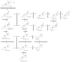
At right is a selection of images that can be regarded as milestones in the development of the steroidogenesis diagram. They are given chronological numbers, with the final version given as number "5" in the results section. Image "4" availed for even easier creation of subsequent works by being a vector image, that is, an image based on a few adjustable points in space rather than millions of pixels. Therefore, subsequent derivatives can easily be edited by free vector graphics editors, in this case Inkscape. Compared to "4", the final version "5" had the following additions:
- Grouping into the major classes of steroids. This step used the textbook Medical Physiology by Boron & Boulpaep[2] as reference, as well as for confirmation of existing molecular structures, enzymes, and products. In addition, for the absence of conversion of corticosterone to cortisol, it used a statement from the Kyoto University Bioinformatics Center that "there is no appreciable conversion of corticosterone to cortisol in the adrenal cortex as 21-OH steroids are poor substrates for 17-alpha hydroxylase."[3] Also, group borders were made transparent between progestagens, mineralocorticoids and glucocorticoids, since many of the included molecules have activities associated with more than one group. For example, cortisol is a typical glucocorticoid, but also has affinity for the mineralocorticoid receptor, although this effect is minor in normal cases because of the enzyme 11β-hydroxysteroid dehydrogenase that breaks down cortisol in the locations where mineralocorticoids play the greatest role.[4] Therefore, the mineralocorticoid field in the diagram also spans over cortisol, but shown in very transparent color because of the small effect. Likewise, corticosterone has strong affinity towards the mineralocorticoid receptor,[5] and it also has an affinity towards the glucocorticoid receptor but this affinity is very weak,[6] and therefore the glucocorticoid field only covers this molecule transparently.
- Addition of enzyme localizations, that is, either in mitochondria or in the smooth endoplasmic reticulum. This information was also taken from Medical Physiology.[2]
- Numbering of the cholesterol carbons, as by convention that is seen for example in the textbook Principles and Practice of Endocrinology and Metabolism.[7]
- Addition of white circles to indicate changes in molecular structure compared with precursors.
- Addition of stereochemistry specifications, that is, standard designations that specify how the atoms are arranged in three dimensions where there are several possible arrangements.
Result
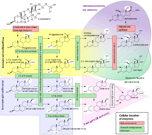
The abbreviation HSD stands for "hydroxysteroid dehydrogenase".
Discussion
This diagram is an example in demonstrating the power of free licensing where anybody can contribute to making things better.
Limitations of the diagram include the fact that there are many additional members of each class. Rather, only the most important pathways are included, in order to provide a model for the creation of the most important steroids, and the main mechanisms by which diseases arise from disturbances in these pathways.
References
- ↑ Hanukoglu I (Dec 1992). "Steroidogenic enzymes: structure, function, and role in regulation of steroid hormone biosynthesis.". J Steroid Biochem Mol Biol 43 (8): 779–804. doi:10.1016/0960-0760(92)90307-5. PMID 22217824.
- 1 2 Walter F. Boron, Emile L. Boulpaep (2003). Medical Physiology: A Cellular And Molecular Approach. Elsevier/Saunders. pp. page 1300. ISBN 1-4160-2328-3.
- ↑ "Steroid hormone biosynthesis - Reference pathway (KO)". Kyoto University Bioinformatics Center. 2013-01-11.
- ↑ Clore, J; Schoolwerth; Watlington, CO (December 1992). "When is cortisol a mineralocorticoid?". Kidney Int. 42 (6): 1297–308. PMID 1474763.
- ↑ Funder, JW. (1997). "Glucocorticoid and mineralocorticoid receptors: biology and clinical relevance.". Annu Rev Med 48: 231-40. doi:10.1146/annurev.med.48.1.231. PMID 9046958.
- ↑ Schaaf, MJ.; Cidlowski, JA. (Mar 2003). "Molecular determinants of glucocorticoid receptor mobility in living cells: the importance of ligand affinity.". Mol Cell Biol 23 (6): 1922-34. doi:10.1128/MCB.23.6.1922-1934.2003. PMID 12612067.
- ↑ Page 705 in:Becker, Kenneth L. (2001). Principles and Practice of Endocrinology and Metabolism. Lippincott Williams & Wilkins. ISBN 978-0-7817-1750-2. http://books.google.com/books?id=FVfzRvaucq8C.