Basic cardiac anatomy
Basic Cardiac Anatomy Level 1: Blood Flow into and out of the Heart
From an anatomical point of view the human heart is composed of four compartments or chambers. An easy way to remember the compartments of the heart is to imagine the heart composed of two apartments like a duplex. There are two halves to the heart separated by a septum (wall). Each compartment is composed of an atrium (the entrance) and a ventricle (living room). When visitors (blood) arrives at either of the two apartments, it enters the atrium through the doors (valves), then moves through to the living room (ventricle) once again through doors (valves).
Dont break my heart, my achey breaky heart JOEY!!!! ill shoot you!!! Blood arrives at the heart through the veins and leaves via the arteries. For example, when blood returns to the heart after travelling around the entire body, it enters through either the superior or the inferior vena cava. When the blood returns to the left atrium after being oxygenated in the lungs, it does so through the pulmonary veins.
The larger of the two chambers is the left side. This results from the greater distance the blood needs to travel once expulsed: after leaving the left ventricle, blood throughout the entire body is pushed. On the otherhand, blood expulsed from the right ventricle only enters the pulmonary circuit -- the lung circuit -- to be reoxygenated. As such, the myocardium of the left side of the heart is vastly more developed than that of the right side.
Roughly speaking, blood circulates in two ways around the body: through pulmonary and systemic circulation. The total circulation follows a figure eight pattern with the heart in the center, between the two loops. The lungs are supplied by the uppermost loop (pulmonary) and the rest of the body is supplied by through the lower loop (systemic). The blood leaves the heart through left atrium, into the aorta (through the aortic valve) and travels to the upper and lower portions of the body. Blood returns to right atrium via the inferior and superior vena cava. From the right atrium, the blood empties into the right heart ventricle through the opening of the tricuspid valve; the blood leaves the right ventricle through the pulmonary semilunar valve and travels through the pulmonary arteries to the left and right lungs where it is reoxygenated. After oxygenation, the blood returns to the left atrium through the pulmonary veins and it passes through the bicuspid (or mitral) valve to the left ventricle. This process is repeated about 60 times per minute, once for each beat of the heart.
Basic Cardiac Anatomy Level 2: Circulation
- 1 Left pulmonary vein
- 2 Right pulmonary vein
- 3 Left atrium
- 4 Mitral valve
- 5 Left ventricle
- 6 Aortic valve (also: Aortic semilunar valve)
- 7 Aorta Ascendens
- 8 Aortic bow
- 9 Aorta Descendens
- 10 Vena Cava Inferior (also: ICV)
- 11 Vena Cava Superior (also: SCV)
- 12 Right atrium
- 13 Tricuspid valve
- 14 Right ventricle
- 15 Pulmonalis valve (also: Pulmonary semilunar valve)
- 16 Truncus pulmonalis
- 17 Right pulmonary artery
- 18 Left pulmonary artery
- 19 Interventricular septum
- 20 Interatrial septum
Note: The major sidebranches of the aorta (around the aortic bow) have not been shown. Many important vessels (arteries supplying the head, neck, and upper limbs) originate in the aortic bow.
We have learned that the heart consists of two atria (singular atrium) and two ventricula (in English, ventricles or the singular, ventricle.)and that blood circulates around the body in two distinct ways. We will now follow the route blood takes when it travels through the heart, beginning with the blood from the lungs.
- 1. Blood is oxygenated in the lungs. Waste materials (Carbon dioxide, CO2) are released and Oxygen, O2 is absorbed by the RBCs (Red Blood Cells).
- 2. The oxygenated blood now travels through the pulmonary veins (venae pulmonales) to the left atrium.
- 3. During the diastolic phase (Diastole: relaxation of the heart), the blood enters the left ventricle through the mitral valve.
- 4. During the systolic phase, (Systole: Contraction of the heart), the heart muscle contracts and the blood is pushed into the aorta ascendens (ascending aorta) through the aortic valve.
- 5. From the aortic bow and the aorta descendens (descending aorta), the blood is spread to smaller arteries which supply every part of the body with fresh oxygenated blood.
- 6. Blood passes through capillaries (hair vessels) where gas exchange between the tissues and the RBCs takes place. The RBCs release oxygen into the tissues and absorb carbon dioxide which will be released again in the lungs.
- 7. The blood leaves the capillaries and flows back into veins.
- 8. The veins combine in the vena cava superior (upper body, above the heart) or vena cava inferior (lower body, under the heart).
- 9. The vena cava superior and inferior both end in the right atrium.
- 10. During diastole, the blood enters the right ventricle through the tricuspid valve.
- 11. During systole, the blood is pushed into the main pulmonary artery (truncus pulmonalis) through the pulmonalis valve.
- 12. Via the lung arteries, the blood reaches the lung capillaries where gas exchange between air and RBCs takes place.
- 13. Back to step 1.
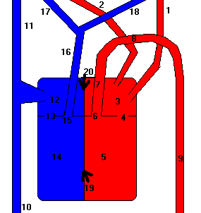
Basic Cardiac Anatomy level 3: Blood Supply of the heart
- MB = Main Branch
- LRAD = Left Ramus Anterior Descendens
- Cx = Circumflex artery
- RCA = Right Coronary Artery
- RDP = Ramus Descendens Posterior
Note: Only the most basic arteries comprising the coronary arterial system are shown here. There are many small branches to each of these arteries; this is only a basic description of the coronary system.
The heart is always working and therefore, needs a continuous uninterrupted supply of fresh oxygenated blood. A system of vessels surrounding the heart, known as the coronary arteries (or simply the coronaries) supply the heart with all the blood it needs. The coronary arteries originate from the aorta ascendens just above the aortic valve. There are two coronaries, one of which splits into two separate coronaries just after its origin; in fact, we could also say that we have three coronaries.
- On the right side, the right coronary artery, runs through the line known as sulcus coronarius (which lies approximately above the border between the right atrium and ventricle) all the way until the back of the heart where it ends in the long interventricular line. Its latter half is known as the Ramus Descendens Posterior. A few centimeters after its origin in the aorta ascendens, the right coronary has a small sidebranch known as the sinus node artery, this supplies the sinus node with blood, and after that, another small branch supplies the front side of the right ventricle with blood.
- The left side of the heart, is more muscular since it needs to push the blood with greater force into the aorta ascendens than the right ventricle needs to push blood into the truncus pulmonalis. This is because the left side of the heart needs to supply every part of the body with blood, from the brain to the fingers and toes.
Basic Cardiac Anatomy level 4: Electrical Activity of the Heart
The pumping action of the heart is initiated by electrical stimulation of the right atrium, by a small pocket of tissue called the Sino-Atrial (SA) node. From here, electrical current follows a conduction pathway through to the ventricles. From the SA node, current travels through both atrium, stimulating their contraction, before coming to the Atrio-Ventricular (AV ) node at the junction of the atria and ventricles. Here, conduction is slowed to allow proper contraction of the atrium (and thus adequate filling of the ventricles with blood). From the AV node, curent flows to the bundle of His (located just below the AV node). It then flows down the Left and Right Bundle Branches, on either side of the intraventricular septum, and to the Purkinje fibres in the ventricules, which cause ventricular contraction.