Vitreous hemorrhage
Background
- Bleeding into the vitreous humor of the eye
- Vitreous is avascular substance that helps keep retina in place
- Traction at its attachments at the ora serrata and optic disc can result in bleeding
- Neovascularization (associated with DM) can result in weak vessels with high propensity for bleeding
- May cause permanent blindness
Causes
- Diabetic retinopathy
- Trauma
- Sickle cell disease
- Posterior vitreous detachment
- Elderly
- Retinal tear
- Terson Syndrome (Association with Subarachnoid Hemorrhage (SAH))
Clinical Features
- Sudden, painless vision loss
- Acute Onset Flashers and Floaters
- Generalized unilateral hazy vision
Differential Diagnosis
Acute Vision Loss (Noninflamed)
- Arteritic anterior ischemic optic neuropathy
- Amaurosis fugax
- Central retinal artery occlusion (CRAO)†
- Central retinal vein occlusion (CRVO)†
- High altitude retinopathy
- Open-angle glaucoma
- Optic neuritis
- Posterior Reversible Encephalopathy Syndrome (PRES)
- Retinal detachment†
- Temporal arteritis†
- Traumatic optic neuropathy
- Vitreous hemorrhage
- Stroke†
†Emergent Diagnosis
Evaluation
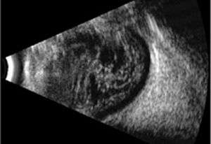
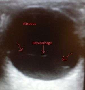
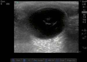
Vitreous Hemorrhage on ultrasound
In the setting of trauma, must assess for Globe Rupture
- Visual acuity
- Degree of vision loss proportional to size of hemorrhage
- Assess for coagulopathy
- INR for patients on warfarin
- Fundoscopy
- May show gross hemorrhage
- Blood may obscure retina
- Decreased red reflex
- Ultrasound
- Bright echoes in posterior chamber
- Small dots or mobile lines may represent early, mild hemorrhage
- Look for retinal injury/tears
- require operative intervention
Management
- Correct coagulopathy
- Ophtho consult (should see ophtho within 24-48 hours)
- Treatment directed at underlying cause
- Avoid NSAIDs and anticoagulants
- Elevate the head of the bed
- Treat nausea/vomiting
References
This article is issued from
Wikem.
The text is licensed under Creative
Commons - Attribution - Sharealike.
Additional terms may apply for the media files.