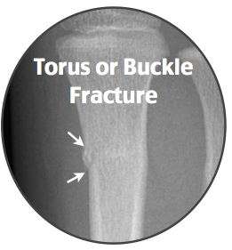
Torus fracture
Background
- Compressive force leads to bulging of the periosteum/cortex
- Also known as buckle fracture
- Often occur at the end of long bones
Clinical Features
- Frequently involves distal radial metaphysis
- Minimal visual deformity
- Soft tissue swelling and point tenderness at injury
Differential Diagnosis
- Greenstick fracture
- Corner fracture (Bucket Handle)
Evaluation
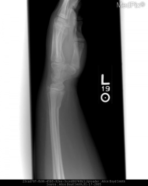
Compression of the cortex and the metadiaphyseal junction consistent with a torus fracture
- Soft tissue swelling and point tenderness
- Visible deformity is unusual
Management
Disposition
- Follow up with pediatrician in 1 week
References
- Geiderman JM, Katz D: General Principles of Orthopedic Injuries, in Marx JA, Hockberger RS, Walls RM, et al (eds): Rosen’s Emergency Medicine: Concepts and Clinical Practice, ed 7. St. Louis, Mosby, Inc., 2010, (Ch) 46:p 473-474.
- Hopkins-Mann C, Ogunnaike-joseph D, Moro-Sutherland D: Musculoskeletal Disorders in Children, in Tintinalli JE, Stapczynski JS, Ma OJ, et al (eds): Tintinalli’s Emergency Medicine, ed 7. New York, The McGraw-Hill Companies Inc., 2011, (Ch) 133
- Koelink, Eric, et al. “Primary Care Physician Follow-up of Distal Radius Buckle Fractures.” Pediatrics, vol. 137, no. 1, 2015, doi:10.1542/peds.2015-2262.
This article is issued from
Wikem.
The text is licensed under Creative
Commons - Attribution - Sharealike.
Additional terms may apply for the media files.