Acute dyspnea
Background
Clinical Features
Emergent Pattern Recognition
| Diagnosis | Lungs | CXR | ECG | Treatment | Contraindicated |
| Pulmonary Edema | Bilateral rales | Interstitial fluid | Normal/abnormal | R/O AMI, lasix, nitrates, ACEi, BiPAP | IVF; ?albuterol; ?Beta-blockers |
| Bronchoconstriction | Wheezes | Clear/hyperinflated | Normal/pulmonary strain | Albuterol, atrovent, steroids, consider anaphylaxis (epi) | Beta-blockers; ?aspirin |
| Pneumonia | Focal ronchi/decreased breath sounds | Infiltrate/effusion | Normal | IVF, antibiotics | Rate control; diuresis |
| Pulmonary embolism | Clear | Clear (most) or Westrmark/Hampton hump | Normal/S1Q3T3 | Anticoagulate/thrombolytics | Rate control |
| Pneumothorax/Hemothorax | Unequal | Pneumo/hemo | Normal | Needle thoracentesis/chest tube | Rate control |
| Dysrythmia | Clear/pulmonary edema | Clear/pulmonary edema | Abnormal | Type dependent | Albuterol; ?IVF |
| ACS | Clear/pulmonary edema | Clear/pulmonary edema | Normal/abnormal | Aspirin; nitrates, anticoagulation, ?beta-blockers, +/- thrombolytics | Albuterol; ?IVF |
Differential Diagnosis
See also Shortness of breath (peds) for pediatric-specific differential
Emergent
- Pulmonary
- Airway obstruction
- Anaphylaxis
- Angioedema
- Aspiration
- Asthma
- Cor pulmonale
- Inhalation exposure
- Noncardiogenic pulmonary edema
- Pneumonia
- Pneumocystis Pneumonia (PCP)
- Pulmonary embolism
- Pulmonary hypertension
- Tension pneumothorax
- Idiopathic pulmonary fibrosis acute exacerbation
- Cystic fibrosis exacerbation
- Cardiac
- Other Associated with Normal/↑ Respiratory Effort
- Abdominal distension
- Anemia
- CO Poisoning
- Salicylate toxicity
- Diabetic ketoacidosis (DKA)
- Diaphragm injury
- Electrolyte abnormalities
- Epiglottitis
- Flail chest
- Hypotension
- Metabolic acidosis
- Pneumonia
- Pneumothorax/hemothorax
- Renal Failure
- Sepsis
- Toxic ingestion
- Other Associated with ↓ Respiratory Effort
Pediatric-specific
- Aspirated foreign body
- Respiratory distress syndrome
- Meconium aspiration syndrome
- Bronchiolitis (peds)
- Pertussis
- Bronchopulmonary dysplasia
- Croup
- Bacterial tracheitis
- Tracheomalacia
- Congenital heart disease
- Vascular ring
- Neonatal abstinence syndrome
- Inborn errors of metabolism
- Brief resolved unexplained event
- Normal neonatal periodic breathing (misinterpreted by caregivers as abnormal)
Non-Emergent
- ALS
- Ascites
- Uncorrected ASD
- Congenital heart disease
- COPD exacerbation
- Fever
- Hyperventilation
- Interstitial lung disease
- Neoplasm
- Obesity
- Panic attack
- Pleural effusion
- Polymyositis
- Porphyria
- Pregnancy
- Rib fracture
- Spontaneous pneumothorax
- Thyroid Disease
- URI
Evaluation
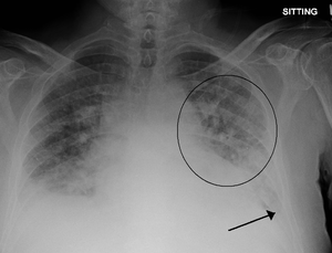
Pulmonary edema with small pleural effusions on both sides.
Bedside Lung Ultrasound in Emergency (BLUE) Protocol[1]
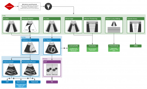
Algorithm for the Use of Ultrasound in the Evaluation of Dyspnea
- Predominant A lines + lung sliding = Asthma/COPD
- Multiple predominant B lines anteriorly with lung sliding = Pulmonary Edema
- Normal anterior profile + DVT= PE
- Anterior absent lung sliding + A lines + lung point = Pneumothorax (PTX)
- Anterior alveolar consolidations, anterior diffuse B lines with abolished lung sliding, anterior asymmetric interstitial patterns, posterior consolidations or effusions with out anterior diffuse B lines = Pneumonia
Management
- Oxygen
- Treat underlying cause
Disposition
See Also
Video
START_WIDGET87ec7f89100d137d-0END_WIDGET
References
- http://ccm.anest.ufl.edu/files/2012/08/BLUELung.pdf Relevance of Lung Ultrasound in the Diagnosis of Acute Respiratory Failure - The BLUE Protocol
This article is issued from
Wikem.
The text is licensed under Creative
Commons - Attribution - Sharealike.
Additional terms may apply for the media files.