Scleritis
Background
- Potentially blinding disorder
- Sclera fuses with dura mater and arachnoid sheath of the opic nerve
- Reason why optic nerve edema and visual compromise are common complications
- 50% of cases associated with an underlying disorder:
Clinical Features
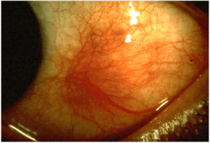
Non-mobile inflammation of entire scleral thickness
- Essential sign is scleral edema, usually accompanied by violaceous discoloration of the globe
- Intense ocular pain that radiates to the face
- Pain with EOM (extraocular muscles insert into the sclera)
- Photophobia
- Globe tenderness to palpation
- Episcleral vessel dilation
Posterior Scleritis
- Posterior to the insertion of the extraocular muscles
- Physical exam often benign
- Inflammation may sometimes be seen at the extremes of gaze
- Patient complains of pain, pain upon EOM
- Involvement of the optic nerve and retina is common
- Retinal detachment, optic disc edema
Complications
- Cornea (peripheral ulcerative keratitis → irreversible loss of vision)
- Uveal tract (anterior uveitis seen in 40% - spillover of inflammation from the sclera)
- Posterior segment (retinal detachment, optic disc edema)
DifferentialDiagnosis
Unilateral red eye
- Acute angle-closure glaucoma^
- Anterior uveitis
- Conjunctivitis
- Corneal erosion
- Corneal ulcer^
- Endophthalmitis^
- Episcleritis
- Herpes zoster ophthalmicus
- Inflamed pinguecula
- Inflamed pterygium
- Keratoconjunctivitis
- Keratoconus
- Nontraumatic iritis
- Scleritis^
- Subconjunctival hemorrhage
- Orbital trauma
^Emergent diagnoses
^^Critical diagnoses
Evaluation
- Labs (to assess possible associated disease)
- CBC
- Chemistry
- Urinalysis (evaluate for glomerulonephritis)
- ESR, CRP
Imaging
- Ultrasound and CT can show thickening of the sclera
Management
- Systemic therapy with NSAIDs, glucocorticoids, or other immunosuppressive drugs
- NSAIDs
- Indomethacin 25-75mg PO TID
Disposition
- Urgent ophtho consult
See Also
References
This article is issued from
Wikem.
The text is licensed under Creative
Commons - Attribution - Sharealike.
Additional terms may apply for the media files.