Retinal hemorrhage
Background
- Due to retinal capillary rupture, can be due to acute or chronic processes
Causes
- Subacute bacterial endocarditis
- Hypertensive retinopathy
- Diabetic retinopathy
- Trauma: nonaccidental trauma (shaken baby syndrome), intracranial hemorrhage, neonatal birth trauma
- Central Retinal Artery Occlusion (CRAO), Central Retinal Vein Occlusion (CRVO)
- Anemia, leukemia, sickle cell anemia
- Anoxia
- Acute mountain sickness
- Carbon monoxide poisoning
- Prolonged intubation during anesthesia
- Connective tissue disease, SLE
- Scurvy, Wernicke-Korsakoff syndrome
- Preeclampsia
- Pentoxifylline
- Ocular decompression following trabeculectomy
Clinical Features
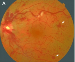
Roth spots due to retinal vein occlusion
- Roth Spots: retinal hemorrhages with white center, seen on fundoscopy
- May be asymptomatic, or cause visual loss
Differential Diagnosis
Acute Vision Loss (Noninflamed)
- Arteritic anterior ischemic optic neuropathy
- Amaurosis fugax
- Central retinal artery occlusion (CRAO)†
- Central retinal vein occlusion (CRVO)†
- High altitude retinopathy
- Open-angle glaucoma
- Optic neuritis
- Posterior Reversible Encephalopathy Syndrome (PRES)
- Retinal detachment†
- Temporal arteritis†
- Traumatic optic neuropathy
- Vitreous hemorrhage
- Stroke†
†Emergent Diagnosis
Evaluation
- Workup dependant on clinical presentation
Management
- Treat underlying condition if appropriate
Disposition
See Also
External Links
References
- Ehlers JP, Shah CP. Wills Eye Manual, The: Office and Emergency Room Diagnosis and Treatment of Eye Disease. 5th ed. Philadelphia, PA:Lippincott Williams & Wilkins; 2008.
- Ling R, James B. White-centred retinal haemorrhages (Roth spots).Postgrad Med J. 1998 Oct;74(876):581-2.
This article is issued from
Wikem.
The text is licensed under Creative
Commons - Attribution - Sharealike.
Additional terms may apply for the media files.