Pyloric stenosis
Background
- More common in males (5:1) & firstborn children (30%)
- Prematurity and macrolide use are also thought to be risk factors
- Most common surgical cause of vomiting in infants
- The main diagnostic criterion is a measurement of more than 3 mm in thickness of the muscular layer.[1]
- Abnormal elongation of the canal is characterised as greater than 17 mm in length .[2][3]
Clinical Features
- Symptoms usually begin between 3-6 weeks of age, rarely after 12 weeks
- Vomiting, immediately postprandial, nonbilious, often projectile, but desires to feed ("hungry vomiter")
- Palpable mass in in RUQ to epigastric region after vomiting, occasionally may see reverse peristaltic fluid wave across abdomen
- If untreated, will see signs of dehydration, weight loss, lethargy, shock
Differential Diagnosis
0–3 Months Old
- Emergent
- Nonemergent
3 mo–3 y old
- Emergent
- Intussusception
- Testicular Torsion
- Trauma
- Volvulus
- Appendicitis
- Toxic megacolon
- Vaso-occlusive crisis
- Nonemergent
3 y old–adolescence
- Emergent
- Appendicitis
- DKA
- Vaso-occlusive crisis
- Toxic ingestion
- Testicular Torsion
- Ovarian Torsion
- Ectopic Pregnancy
- Trauma
- Toxic megacolon
- Inflammatory bowel disease
- Gastric ulcer disease
- Ovarian cyst
- Pregnancy
- Pancreatitis
- Cholecystitis
- Intussusception (to age 6)
- Nonemergent
Evaluation
- Labs may show hypokalemia and hypochloremic metabolic alkalosis
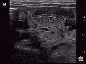
Longitudinal view of thickened and elongated pylorus muscle[4]
Imaging
Primary test of choice
- Ultrasound: thickened (>3 mm) and elongated (>17 mm) pylorus ~ 95% Sn/Sp[5]
Other tests that may show findings
- Abdominal x-ray: may show large stomach bubble with absence of air in small bowel or colon
- Characteristic caterpillar sign (gastric contractions against hypertrophied pylorus)
- Upper GI: string sign (narrowed pyloric lumen), double track sign (duplicated mucosa), beak sign (abnormality of pyloric opening)
Management
- IVF
- Normal electrolytes and no evidence of dehydration
- 5% dextrose with 0.25% NaCl and 2 meq KCl per 100 mL
- Moderate or severe dehydration
- Higher NaCl concentrations (0.5% to normal saline) and higher rates of administration (1.5 to 2 times maintenance)
- Ensure correction of bicarbonate level, as it may be a hypoventilation risk
- Normal electrolytes and no evidence of dehydration
- Nasogastric tube
- Surgery
- Can be delayed 24-36 hr to rehydrate infant and correct electrolytes
Precautions
- Ensure that kidneys are functional prior to giving potassium
- Do not give lactated ringers
- May lead to worsening alkalosis > apnea in infants
Disposition
- Admission
See Also
References
- Dias, S et al Insights Imaging. 2012 Jun; 3(3): 247–250.
- Teele RL, Smith EH. Ultrasound in the diagnosis of idiopathic hypertrophic pyloric stenosis. N Engl J Med. 1977 May 19. 296(20):1149-50.
- Sargent SK, Foote SL, Mooney DP, Shorter NA. The posterior approach to pyloric sonography. Pediatr Radiol. 2000 Apr. 30(4):256-7
- http://www.thepocusatlas.com/pediatrics/
- Rohrschneider WK, Mittnacht H, Darge K, Tröger J. Pyloric muscle in asymptomatic infants: sonographic evaluation and discrimination from idiopathic hypertrophic pyloric stenosis. Pediatr Radiol. 1998 Jun;28(6):429-34.
This article is issued from
Wikem.
The text is licensed under Creative
Commons - Attribution - Sharealike.
Additional terms may apply for the media files.