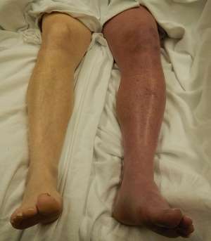
Phlegmasia cerulea dolens
Background
- "Painful Blue Leg"
- Massive iliofemoral occlusion due to venous thromboembolism
- Extensive vascular congestion and venous ischemia
Clinical Features
Two hour history of phlegmasia cerulea dolens (left leg)
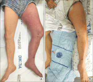
At presentation (A) and post-op day 1 (B).
- Sudden severe leg pain
- Swelling and edema (bleb/bullae)
- Cyanosis
- Venous gangrene
- Compartment syndrome
- Arterial compromise
- Shock
- Often preceded by phlegmasia alba dolens
Differential Diagnosis
Clinical Spectrum of Venous thromboembolism
- Deep venous thrombosis (uncomplicated)
- Phlegmasia alba dolens
- Phlegmasia cerulea dolens
- Venous gangrene
- Pulmonary embolism
- Isolated distal deep venous thrombosis
Only 40% of ambulatory ED patients with PE have concomitant DVT[1][2]
- Cellulitis
- Lymphedema
- Venous valvular insufficiency
- Superficial thrombophlebitis
Evaluation
- Clinical diagnosis
- Duplex US
- Contrast venography
Management
- For mild, non-gangrenous form: Conservative management
- Steep limb elevation
- Fluid resuscitation
- Heparin: 80-100U/kg followed by infusion of 15-18U/kg/hr
- Vascular surgery consult for emergent thrombectomy
- Interventional radiology consult for emergent catheter-directed thrombolysis
- Thrombolytic therapy: Alteplase (1mg/min to total of 50mg) distal to thrombus
Disposition
- Admit
See Also
External Links
References
- Righini M, Le GG, Aujesky D, et al. Diagnosis of pulmonary embolism by multidetector CT alone or combined with venous ultrasonography of the leg: a randomised non-inferiority trial. Lancet. 2008; 371(9621):1343-1352.
- Daniel KR, Jackson RE, Kline JA. Utility of the lower extremity venous ultrasound in the diagnosis and exclusion of pulmonary embolism in outpatients. Ann Emerg Med. 2000; 35(6):547-554.
This article is issued from
Wikem.
The text is licensed under Creative
Commons - Attribution - Sharealike.
Additional terms may apply for the media files.