Nailbed laceration
Background
- Results from a crush injury or blunt trauma
Nailtip Anatomy
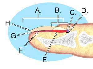
A. Nail plate; B. lunula; C. root; D. sinus; E. matrix; F. nail bed; G. hyponychium; H. free margin.
- The perinychium includes the nail, the nailbed, and the surrounding tissue.
- The paronychia is the lateral nail folds
- The hyponychium is the palmar surface skin distal to the nail.
- The lunula is that white semi-moon shaped proximal portion of the nail.
- The sterile matrix is deep to the nail, adheres to it and is distal to the lunule.
- The germinal portion is proximal to the matrix and is responsible for nail growth.
Clinical Features
- Laceration of the nail bed
- May also include nail avulsion and/or distal phalanx fracture
Differential Diagnosis
Evaluation
- Plain films of the involved digits to evaluate for fracture
Management
- Remove overlying nail, if present
- Repair lacerations using 5-0 or 6-0 absorbable sutures
- Replace nail into nail fold
- Trephination of the nail may be performed to allow drainage of blood
- Nail may be sutured into place
- Alternatively, a nail-shaped adaptic or non-adherent gauze may be placed under the nail fold
Prognosis
- Complete nail regrowth may take 70 to 160 days
- Potential risk of nail deformity and losing the nail
Disposition
- Discharge
External Links
See Also
This article is issued from
Wikem.
The text is licensed under Creative
Commons - Attribution - Sharealike.
Additional terms may apply for the media files.