Melanoma
Background
- Ultraviolet UVB light (315 – 280 nm) from sun/tanning beds damages DNA
- High risk in fair skin, blue eyes, and red-haired people,
- High risk history of multiple atypical nevi or dysplastic nevi
Clinical Features
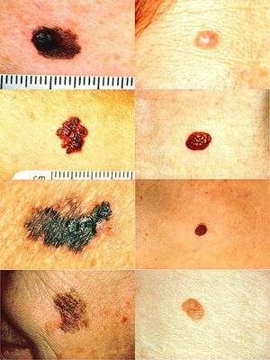
On the left side from top to bottom: melanomas showing (A) Asymmetry, (B) a border that is uneven, ragged, or notched, (C) coloring of different shades of brown, black, or tan and (D) diameter that had changed in size compared to normal moles on the right.
- Asymmetry
- Borders (irregular)
- Color (variegated)
- Diameter (greater than 6 mm (0.24 in), about the size of a pencil eraser)
- Evolving over time or Elevated above the skin
Differential Diagnosis
Evaluation
- Biopsy outpatient at dermatology office
- LDH (elevated indicates metastasis, particularly to the liver, not sensitive)
- Imaging for metastasis
- CXR or CT
- MRI, PET and/or PET/CT scans
Disposition
- Refer to dermatology
Prevention
- Avoid UV radiation: hats, long sleeve shirts, sunglasses with UV protection
- Even though tanning beds mostly UVA, still can cause melanoma
- Sunscreen
- Blocks damaging UVB radiation
- Sun Protectant Factor (SPF) of 15 means if you notice a burn at 10 min, applying SPF 15 would allow you to stay out 150 min before noticing burn
- SPF >30 if outside work or long time recreation and reapply as needed
See Also
References
This article is issued from
Wikem.
The text is licensed under Creative
Commons - Attribution - Sharealike.
Additional terms may apply for the media files.