ECG axis
ECG Axis
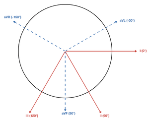
Limb and augmented leads
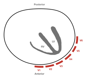
Precordial leads
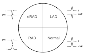
Normal Axis
- -30°→90°
- Dominant QRS direction (positive or negative) can be used to approximate axis
- Normal axis if positive QRS in leads I and aVF
Right Axis Deviation
- Causes:
- Usually accompanied by tall R wave in V1
- Right ventricular hypertrophy
- Pulmonary hypertension & chronic lung disease
- COPD
- Pulmonary embolism
- Left posterior fascicular block
- Lateral MI (from Q-waves in lead I)
- Ventricular ectopy (VT)
- TCA toxicity, sodium channel blocker toxicity
- Hyperkalemia
- Lead misplacement
- Dextrocardia
- Normal thin adults with horizontally positioned hearts
- Usually accompanied by tall R wave in V1
Left Axis Deviation
- Causes:
- Left anterior fascicular block
- Left Bundle Branch Block
- Inferior MI (from Qs)
- Left Ventricular Hypertrophy
- Pacer
- WPW
- Hyperkalemia
- Normal variant
See Also
Video
START_WIDGETc0207b6806a0b4b8-0END_WIDGET
External Links
References
This article is issued from
Wikem.
The text is licensed under Creative
Commons - Attribution - Sharealike.
Additional terms may apply for the media files.