Le Fort fractures
Background
- Midface fracture involving the maxilla and surrounding facial structures
- Most commonly occur due to motor vehicle accident
- LeFort I fractures are isolated to the lower face
- Type II and III injuries associated with cribriform plate disruption and CSF rhinorrhea
Clinical Features
Differential Diagnosis
Evaluation
Workup
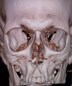
A 3-D CT reconstruction showing a Le Fort type 1 fracture (marked by arrow).
- CT sinus/face
Diagnosis
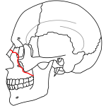
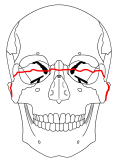
| Le Fort Fracture | Description | Front View | Side View | Stability |
| Type I |
|
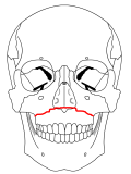 | 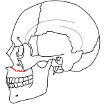 | Stable |
| Type II |
|
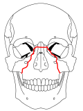 |  | Can be stable or unstable fracture |
| Type III |
|
 | 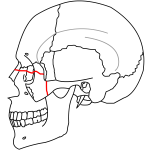 | Unstable |
| Type IV |
|
Unstable |
Management
- Ensure airway patency
- If intubation required, consider awake intubation
- Control hemorrhage
- Nasal and oral packing may be required
- IV antibiotics
Disposition
- Consider discharge in isolated LeFort I or stable LeFort II fractures without concerning features (in coordination with appropriate specialist consult - OMFS, ENT, or PRS)
- All others should be admitted
See Also
References
- Tintinalli 7th Edition, pgs 1730-1738
This article is issued from
Wikem.
The text is licensed under Creative
Commons - Attribution - Sharealike.
Additional terms may apply for the media files.