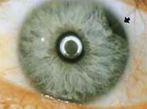
Iridodialysis
Background
- Localized separation of the iris from the ciliary body
- Most commonly caused by blunt trauma to the eye[1]
- May also be caused by penetrating eye trauma or as an iatrogenic injury during ophthalmologic procedures
Iridodialysis
Differential Diagnosis
Orbital trauma
Acute
- Caustic keratoconjunctivitis^^
- Conjunctival laceration
- Corneal abrasion
- Globe rupture^
- Iridodialysis
- Ocular foreign body
- Orbital fracture
- Frontal sinus fracture
- Naso-ethmoid fracture
- Inferior orbial wall fracture
- Medial orbital wall fracture
- Retinal detachment
- Retrobulbar hemorrhage/hematoma
- Subconjunctival hemorrhage
- Traumatic hyphema
- Traumatic iritis
- Traumatic mydriasis
Subacute/Delayed
Evaluation
- Clinical diagnosis
Management
- Simple iridodialysis requires no specific ED treatment
- Small iridodialysis often managed conservatively - if large and/or symptomatic, generally requires surgical repair[1]
- If associated with hyphema, see hyphema management
Disposition
- Based on discussion with ophthalmology
See Also
External Links
References
- Pandav, S. S., Gupta, P. C., Singh, R. R., Das, K., Kaushik, S., Raj, S., & Ram, J. (2016). Cobbler’s Technique for Iridodialysis Repair. Middle East African Journal of Ophthalmology, 23(1), 142–144. http://doi.org/10.4103/0974-9233.171770
- Omar Yousif, M. (2016). Single suture customized loop for large iridodialysis repair. Clinical Ophthalmology (Auckland, N.Z.), 10, 1883–1890. http://doi.org/10.2147/OPTH.S111322
This article is issued from
Wikem.
The text is licensed under Creative
Commons - Attribution - Sharealike.
Additional terms may apply for the media files.