Intertrochanteric femur fracture
Background
- Occur via fall in elderly or osteoporotic
Clinical Features
- Typically pain, swelling, ecchymosis
- May lose 1-2L of blood
- Unable to bear weight
- Shortening and external rotation if fracture is significantly displaced
Differential Diagnosis
Proximal
- Intracapsular
- Extracapsular
Evaluation
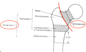
Location of femur fractures
- Consider AP pelvis in addition to AP/lateral views to compare contralateral side
- Consider MRI if strong clinical suspicion but negative x-ray
Evaluation

Garden's classification of intertrochanteric fractures
- Stable (Garden's type I and II)
- Lesser trochanter non-displaced, no comminution, medial cortices of prox/distal fragments aligned
- Unstable (Garden's type III and IV)
- Displacement occurs, comminution is present, or multiple fracture lines exist
Management
General Fracture Management
- Acute pain management
- Open fractures require immediate IV antibiotics and urgent surgical washout
- Neurovascular compromise from fracture requires emergent reduction and/or orthopedic intervention
- Consider risk for compartment syndrome
Specific Management
- Ortho consult
Disposition
- Admit
Specialty Care
- Typically requires ORIF
See Also
External Links
References
This article is issued from
Wikem.
The text is licensed under Creative
Commons - Attribution - Sharealike.
Additional terms may apply for the media files.