Ingrown toenail
Background
Nailtip Anatomy
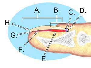
A. Nail plate; B. lunula; C. root; D. sinus; E. matrix; F. nail bed; G. hyponychium; H. free margin.
- The perinychium includes the nail, the nailbed, and the surrounding tissue.
- The paronychia is the lateral nail folds
- The hyponychium is the palmar surface skin distal to the nail.
- The lunula is that white semi-moon shaped proximal portion of the nail.
- The sterile matrix is deep to the nail, adheres to it and is distal to the lunule.
- The germinal portion is proximal to the matrix and is responsible for nail growth.
- Lateral nail edge grows deep into nail wall → cycle of inflammation and hypertrophic granulation tissue can lead to abscess
Clinical Features
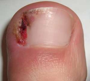
Typical ingrown toenail
- Pain and swelling at distal nailbed, typically unilateral
Differential Diagnosis
Acute
- Foot and toe fractures
- Subtalar dislocation
- Metatarsophalangeal sprain (turf toe)
- Acute arterial ischemia
- Calcaneal bursitis
Subacute/Chronic
- Diabetic foot infection
- Peripheral artery disease
- Plantar fasciitis
- Trench foot
- Ingrown toenail
- Paronychia
- Tinea pedis
- Morton's neuroma
- Diabetic neuropathy
Evaluation
Workup
- Typically does not require studies
Diagnosis
- Typically a clinical diagnosis
Management
- Minor cases can be treated non-surgically
- Others, preform partial ingrown toenail removal
Disposition
- Outpatient
See Also
External Links
References
This article is issued from
Wikem.
The text is licensed under Creative
Commons - Attribution - Sharealike.
Additional terms may apply for the media files.