Femur fracture
This page is for adult patients. For pediatric patients, see: femur fracture (peds)
Background
- Despite good care, proximal fracture 30-day all cause mortality is 22% and grows to 36% at one year[1]
Proximal
- Intracapsular
- Extracapsular
Clinical Features
- History of trauma
- Pain, point tenderness, deformity
Differential Diagnosis
Evaluation
Proximal
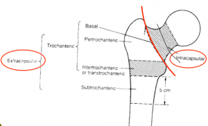
Location of femur fractures
- Consider AP pelvis in addition to AP/lateral views to compare contralateral side
- Consider MRI if strong clinical suspicion but negative x-ray
Mid-Shaft
- Plain xrays of femur
Management
General Fracture Management
- Acute pain management
- Open fractures require immediate IV antibiotics and urgent surgical washout
- Neurovascular compromise from fracture requires emergent reduction and/or orthopedic intervention
- Consider risk for compartment syndrome
Specific Management
- Pain control in ED with femoral nerve block
- Nerve Block: Fascia Iliaca Compartment
- 3 in 1 block (femoral, obturator, lateral cutaneous nerve of thigh)
- No difference in 2 blocks listed above, which both reduced pain scores in the ED. [2]
- Type and cross/screen for patients at higher risk of hemorrhage:
- Age > 75 yrs
- Initial hemoglobin < 12
- Peritrochanteric fracture
Disposition
- Generally requires admission for operative repair
Specialty Care
- Most fractures, including all displaced, are treated with ORIF
- Exception is isolated trochanteric fracture often does not require surgery
- See individual pages for further discussion
See Also
References
- Lawrence, VA, et al. Medical complications and outcomes after hip fracture repair. Arch Intern Med. 2002; 162(18):2053-7.
- Reavley P, et al. Randomised trial of the fascia iliaca block versus the ‘3-in-1’ block for femoral neck fractures in the emergency department. Emerg Med J. 2014 Nov 27.
This article is issued from
Wikem.
The text is licensed under Creative
Commons - Attribution - Sharealike.
Additional terms may apply for the media files.