FAST exam
Background
- "Focused assessment with sonography for trauma"
- Prioritize: Do primary survey of ATLS first (ABCDE)
- FAST exam follows ABCDE to assess “C” looking for free fluid
- Sensitivity of 42% and specificity of ≥98%[1][2]
- As little as 100 mL of free fluid can be seen[3][4], though >500 mL is needed for the common user[5]
- If blunt trauma start with RUQ view first
- In penetrating start with cardiac views first to rule out tamponade
- Serial exams extremely helpful
Indications
- FAST is useful in patients with blunt or penetrating traumatic injury
- Enables trauma bay decision:
- Stable patient with traumatic mechanism of injury + negative FAST → observation
- Stable patient with traumatic mechanism of injury + positive FAST → CT
- Unstable patient with traumatic mechanism of injury + negative FAST → repeat FAST or CT
- Unstable patient with traumatic mechanism of injury + positive FAST → laparotomy
Technique
- Select probe
- Curvilinear/large convex probe is ideal but phased array probe may be substituted
- Location
- Sequence can vary depending on mechanism of injury
- Include cardiac, RUQ, pelvic, and LUQ views
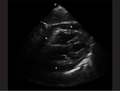
Cardiac
Pericardial fluid on ultrasound
.gif)
Transthoracic echo of pericardial fluid showing "swinging heart"
- Location
- Subxiphoid
- Landmarks
- Visualize the heart and pericardium using the liver as an acoustic window
- Scan anterior to posterior through the heart
RUQ
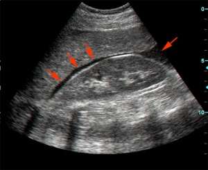
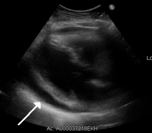
A positive FAST - fluid (black stripe, indicated by red arrows) within Morison's pouch.
- Location
- Coronal view over the right flank
- Landmarks
- Visualize the interface between the liver and kidney
- Scan anterior to posterior identifying Morison’s pouch and the superior and inferior pole of the kidney
Pelvic
- Location
- Sagittal view just superior to the pubic symphysis
- Landmarks
- Identify the bladder
- Scan medial to lateral to identify fluid posterior and superior to the bladder
LUQ
- Location
- Coronal view over the left flank
- Landmarks
- Identify the space between the spleen and diaphragm and the spleen and the kidney
- Scan through anterior to posterior of the splenodiaphragmatic space and superior and inferior pole of the kidney
Findings
- Positive FAST will have one of the following:
- Anechoic area within the pericardial space
- Anechoic areas between the liver and kidney
- Anechoic areas between the diaphragm and spleen
- Anechoic areas between the spleen and kidney
- Anechoic areas between superior and posterior to the posterior wall of the bladder
Images
Normal
.gif) Normal subxiphoid view
Normal subxiphoid view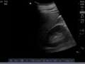 RUQ with no free fluid
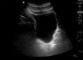 Normal saggital bladder
Abnormal
 Pericardial effusion
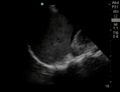 Positive FAST (RUQ)
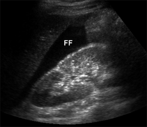 Positive FAST (RUQ)
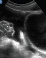 Free fluid superior to the bladder
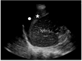 Positive FAST (LUQ)
Pearls and Pitfalls
- Morison’s pouch
- Scans must scan through the inferior poles of the kidneys as this can contain small quantities of fluid
- Serial exam may be needed
- Negative exam does not rule out intraabdominal injuries
- Retroperitoneal hemorrhage not easily identified
- Those with delayed presentation may have clotted and not demonstrate completely anechoic fluid collections
Documentation
Normal Exam
A bedside FAST ultrasound was conducted to assess for free fluid with clinical indication of trauma. Cardiac, RUQ, pelvic, and LUQ views were adequately obtained. There was no free fluid identified.
Abnormal Exam
A bedside FAST ultrasound was conducted to assess for free fluid with clinical indication of trauma. Cardiac, RUQ, pelvic, and LUQ views were adequately obtained. There was free fluid identified in the RUQ suggesting intraabdominal hemorrhage.
Clips
Normal
 Normal subxiphoid view
 Normal pelvic view
Abnormal
 Free fluid located at the liver tip
External Links
See Also
References
- Natarajan B, Gupta PK, Cemaj S, et al. FAST scan: Is it worth doing in hemodynamically stable blunt trauma patients? Surgery. 2010;148(4):695-700.
- Miller MT, Pasquale MD, Bromberg WJ, et al. Not so FAST. J Trauma. 2003; 54(1):52-59.
- Goldberg GG. Evaluation of ascites by ultrasound. Radiology. 1970; 96(15):217–221.
- Von Kuenssberg Jehle D, Stiller G, Wagner D. Sensitivity in detecting free intraperitoneal fluid with the pelvic views of the FAST exam. Am J Emerg Med. 2003 Oct;21(6):476-478.
- McKenney KL, McKenney MG, Cohn SM, et al. Hemoperitoneum score helps determine need for therapeutic laparotomy. J Trauma 2001; 50(4):650–654.
This article is issued from
Wikem.
The text is licensed under Creative
Commons - Attribution - Sharealike.
Additional terms may apply for the media files.