Distal radius fractures
Background
- Definition: Fracture at the metaphysis or the articulation of the distal radius
- One of the most common fractures seen in the ED (1/6th of all fractures treated)
Distal radius fractures
- Colles' fracture
- Smith's fracture
- Barton's fracture
- Radial styloid fracture
- Distal radioulnar joint disruption
Distal radius fracture eponyms
| Eponyms | Description |
| Barton's | Fracture-dislocation of radiocarpal joint (with intra-articular fracture involving the volar or dorsal lip) |
| Chauffer's | Fracture of radial styloid |
| Colles' | Dorsally displaced, extra-articular fracture |
| Die-punch | Depressed fracture of the lunate fossa (articular surface) |
| Smith's | Volar displaced, extra-articular fracture |
Clinical Features
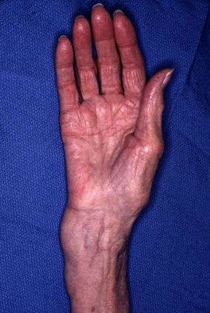
Distal radius fracture demonstrating the deformity in the wrist.
- Commonly from a fall on an outstretched wrist that is dorsiflexed
- Wrist/forearm trauma and pain
- Possible forearm deformity
Exam Pearls
- Perform full neurovascular assessment of the hand (including median, ulnar, and radial nerves
- Examine ipsilateral elbow, shoulder, and hand
Differential Diagnosis
Forearm Fractures
- Distal radius fractures
- Colles' fracture
- Smith fracture
- Barton fracture
- Radial styloid fracture
- Distal radioulnar joint disruption
- Radia ulna fracture
- Isolated radius fracture (proximal)
- Isolated ulna fracture (i.e. nightstick)
- Monteggia fracture-dislocation
- Galeazzi fracture-dislocation
- Forearm fracture (peds)
Evaluation
Workup
- Forearm x-ray AP and lateral
Diagnosis
- Typically from plain forearm x-rays
Management
General Fracture Management
- Acute pain management
- Open fractures require immediate IV antibiotics and urgent surgical washout
- Neurovascular compromise from fracture requires emergent reduction and/or orthopedic intervention
- Consider risk for compartment syndrome
Acute Reduction
- Indications:
- Most angulated and/or displaced distal radius requires closed reduction and placement of a sugar-tong splint
- Consider even if operative management is expected (to reduce pain and swelling)
- Steps:
- Adequate analgesia (e.g. morphine and/or hematoma block)
- highly consider procedural sedation
- Axial traction: Manual or finger traps with hanging weights, if available
- Recreate, then reverse, mechanism of injury
- Although recreating the injury briefly exaggerates the existing deformity, this maneuver "unlocks" any periosteal sleeve folded into the fracture site (which can be critical in achieving reduction)
- Continue to maintain axial traction throughout manuver
- For example with a fracture with dorsally angulated fragments, establish axial traction, then slightly bending the dorsal fragment even more dorsally while maintain traction, then reverse pressure to reduce the distal fragment volarly and back out to length, all while maintaining traction.
- Splint
- Re-image
- Goal:
- Always target optimal (anatomic) fracture reduction
- Acceptable healing parameters:
Operative fixation is recommended for fractures with post-reduction radial shortening > 3mm, dorsal tilt > 10°, or intra-articular displacement or step-off > 2mm[1]
Disposition
- Most can be treated with orthopedic follow up within 1 week
Admit
- Open fracture
- Neuro-vascular compromise
- Risk or concern for compartment syndrome
- Patient is unable to function at home (e.g. uses walker with that arm)
See Also
References
- AAOS OrthoGuidelines. Treatment of Distal Radius Fractures. http://www.orthoguidelines.org/topic?id=1003
This article is issued from
Wikem.
The text is licensed under Creative
Commons - Attribution - Sharealike.
Additional terms may apply for the media files.