Fundoscopic exam
Background
- optic disc for cupping/pallor/swelling/hemorr
- central retina for hemor/pallor
- periph retina for vessel appearance/hemorr/detach
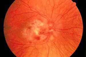
Retinal Images
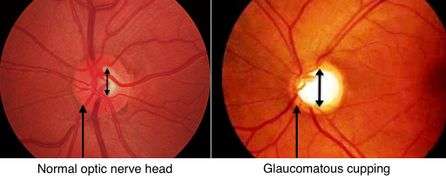 Open-angle glaucoma (cupping)
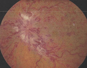
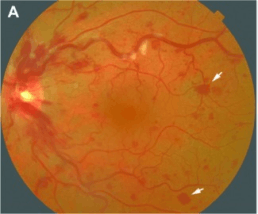 Roth spots due to retinal vein occlusion (retinal hemorrhage)
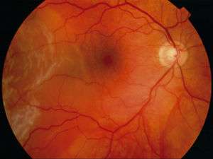
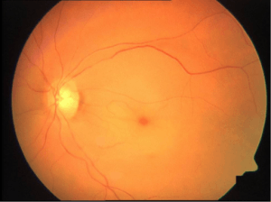 Central retinal artery occlusion: cherry-red spot, retinal edema and narrowing of the vessels.
See Also
This article is issued from
Wikem.
The text is licensed under Creative
Commons - Attribution - Sharealike.
Additional terms may apply for the media files.