Dental subluxation
Background
Clinical Features
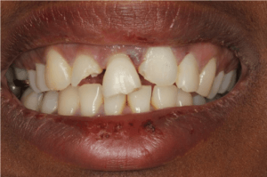
Coronal incisor fractures and with extrusive luxation of the upper right central incisor.
- Tooth is mobile but is still in original anatomic position
- If multiple consecutive teeth are involved, rule out underlying alveolar ridge fracture or other Mandible fracture
Differential Diagnosis
Evaluation
- Clinical diagnosis
Management
Extrusive Luxation
- (tooth is moved partially out of the socket)
- Reposition tooth
- Follow up within 24hr for stabilization
- Temporizing measure: Periodontal pack (e.g.-Coe-Pak) in which tooth is bonded to its two neighboring teeth on both sides
- Mix the resin and catalyst paste and apply to completely dry teeth
- May use nasal cannula with oxygen as a air/drying source
- May place gauze rolls in mucobuccal fold to absorb saliva
- Wet or lubricated goves will allow for easier handling
- Apply splinting to the facial side of the teeth, spanning approximately 1-2 teeth in either direction
- Avoid covering the occlusal (biting) surface
Lateral Luxation
- (tooth displaced in a direction other than inward or outward)
- More extensive injury than extrusive luxation
- Associated with cracking or fracture of the surrounding alveolar bone
- Attempt repositioning of tooth
- Apply temporary splinting with periodontal dressing
- Follow up within 24hr for stabilization
Intrusive Luxation
- (tooth is forced inward into the socket)
- Most serious because of significant damage to alveolar socket and periodontal ligament
- Allow tooth to erupt on its own
Disposition
- Discharge with dental follow up
See Also
- Dental Problems
References
This article is issued from
Wikem.
The text is licensed under Creative
Commons - Attribution - Sharealike.
Additional terms may apply for the media files.