Chest X-ray interpretation
ABC Method
- Airways - ex. FB, trachea deviation
- Bones and soft tissue - ex. Rib fractures, clavicle fracture
- Cardiac - ex. Cardiomegaly
- Diaphragm - ex. Free air
- Effusions - ex. Pleural effusion
- Fields and Fissures - ex. Congestion, consolidations
- Great vessels - ex. Apical cap
- Hila and mediastinum - Widened mediastinum
- Impression
DRSABCDE Method
- Details
- Info of patient
- PA/AP, Lateral
- RIPE - Image quality
- Rotation
- Inspiration
- Picture
- Exposure
- Soft tissues and bones
- Airway & mediastinum
- Breathing
- Lung fields
- Circulation
- Heart position and size
- Diaphragm
- Extras
- Lines, tubes, catheters
Examples
 Pneumoperitoneum
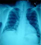
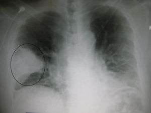 Prominent wedge-shape area of airspace consolidation in the right lung, characteristic of bacterial pneumonia
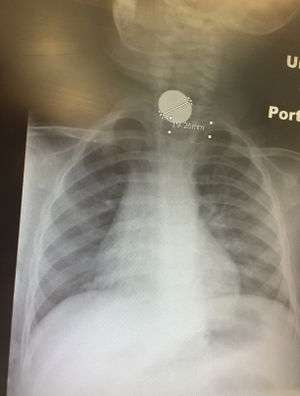 Esophageal foreign body (penny in 12 mo male)
 Right sided pneumothorax
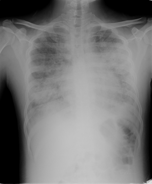
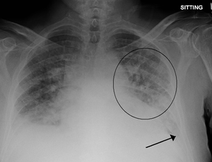 Pulmonary edema with small pleural effusions on both sides.
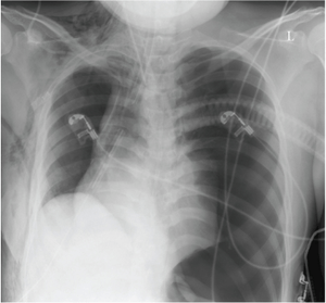 Left sided tension pneumothorax with mediastinal shift
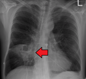
Special Findings
Elevated Hemidiaphram
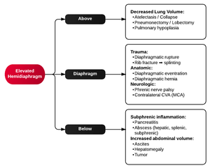
Elevated hemidiaphragm algorithm
See Also
This article is issued from
Wikem.
The text is licensed under Creative
Commons - Attribution - Sharealike.
Additional terms may apply for the media files.