Basilar skull fracture
Background
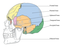
Bones of the cranium.
- Involve at least one of five bones that comprise base of the skull [1]
- cribriform plate of ethmoid bone
- orbital plate of the frontal bone
- petrous and squamous portion of the temporal bone
- sphenoid and occipital bones
- Occur most commonly through temporal bone--> high risk for extra-axial hematomas, particularly epidural hematoma
Clinical Features
.jpg)
Raccoon eyes
- Nausea/vomiting, oculomotor deficits from injuries to CN3, 4 or 6
- Retroauricular or mastoid ecchymosis (Battle sign), onset 1-3 days after fracture occurred
- Raccoon eyes: periorbital ecchymosis
- Clear rhinorrhea or otorrhea
- "halo" sign: drop of fluid placed on tissue or filter paper, rapidly expanding ring of clear fluid around red blood defines positive test
- CSF distinguished from local nasal secretions with presence of beta-trace protein or beta-2 transferrin
- Hemotympanum
Differential Diagnosis
Head trauma
- Traumatic brain injury
- Orbital trauma
- Maxillofacial trauma
- Skull fracture
- Pediatric head trauma
Evaluation
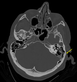
Temporal bone fracture as seen on CT.
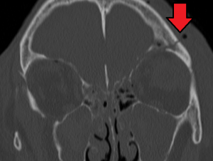
CT showing basilar skull fracture
- Noncontrast CT head
Management
- Immediate neurosurgical consultation
Disposition
- Admit for observation regardless of need for surgical intervention
See Also
- Skull Fracture
External Links
- Pillai, Saran S. Basilar Skull Fracture: Basics & Beyond available at http://www.emdocs.net/basilar-skull-fracture-basics-beyond/
References
- Golfinos JG, Cooper PR. Skull fracture and post-traumatic cerebrospinal fluid fistula. In:Head Injury, 4th, Cooper PR, Golfinos JG (Eds), McGraw-Hill, New York 2000. p.155
This article is issued from
Wikem.
The text is licensed under Creative
Commons - Attribution - Sharealike.
Additional terms may apply for the media files.