Ankle dislocation
Background
- Most ankle dislocations are associated with a fracture
- Must rule-out neurovascular compromise and conversion to open fracture
- Reduce immediately if vascular compromise or skin tenting is present
- Posterior dislocation is most common
- Assoc with rupture of tibiofibular ligaments or lateral malleolus fracture
Clinical Features
- Ankle pain/trauma/deformity
Evaluation
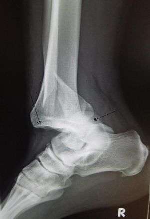
Rraumatic dislocation of the ankle (tibiotarsal) with distal fibular fracture. Open arrow marks the tibia and the closed arrow marks the talus.
- Ankle x-ray
Management
Posterior dislocation [1]
- Assistant places hands under knee and distal thigh to pull counter traction
- Hold dorsum of mid foot with one hand and heel with other hand. Pull longidtudinally then anteriorly
- If no assistant, have patient hang leg over edge of stretcher
Anterior dislocation
- As above, but dorsiflex foot first to disengage talus
- Then axial traction while assistant is holding traction on tibia
- Finally push foot posteriorly while assistant adds pulls anteriorly
Lateral dislocation
- Plantar flex foot then apply traction with assistant holding counter traction
Post reduction (all)
- Check pulses after any attempts. If not palpable, consult ortho emergently
- Document pulse/motor/sensory exam before and after any attempts at reduction
- Splint in posterior as well as sugar tong splint with foot in 90 degree flexion
- Flex hip and knee to 90 degrees to relax gastroc/soleus
Disposition
External Links
References
- Davenport M. Procedures for orthopedic emergencies. In: Bond M, ed. Orthopedic Emergencies: Expert Management for the Emergency Physician. Cambridge: Cambridge University Press; October 31, 2013.
This article is issued from
Wikem.
The text is licensed under Creative
Commons - Attribution - Sharealike.
Additional terms may apply for the media files.