Acute respiratory distress syndrome
Background
- Non-cardiogenic pulmonary edema due to lung capillary endothelial injury (diffuse alveolar damage)
- Proteinaceous material accumulate in alveoli in a heterogeneous manner (hyalinosis)
- Symptom of an underlying disease
Clinical Features
Diagnostic Criteria[1]
- New onset respiratory symptoms
- Bilateral pulmonary opacities
- Symptoms not explained by cardiac etiology or volume overload
Severity by Berlin Definition[1]
| PaO2/FiO2 | Severity | Mortality |
| 200-300 | Mild | 27% |
| 100-200 | Mod | 32% |
| <100 | Severe | 45% |
Imaging
- Diffuse patchy pulmonary infiltrates
Differential Diagnosis
Pulmonary Edema Types
Noncardiogenic pulmonary edema
- Negative pressure pulmonary edema
- Upper airway obstruction
- Reexpansion pulmonary edema
- Strangulation
- Neurogenic causes
- Iatrogenic fluid overload
- Multiple blood transfusions
- IV fluid
- Inhalation injury
- Pulmonary contusion
- Aspiration pneumonia and pneumonitis
- Other
- High altitude pulmonary edema
- Hypertensive emergency
- ARDS
- Sympathetic crashing acute pulmonary edema (SCAPE)
- Immersion pulmonary edema
- Hantavirus pulmonary syndrome
- Missed dialysis in kidney failure
Evaluation
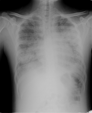
ARDS on CXR
Management
- Treat underlying cause
- Supplemental O2
- Noninvasive ventilation
- Limited data to support use
- Consider pulse dose steroids in early, established, severe ARDS in ICU setting[3]
- NOT for prevention of ARDS (will increase risk for ARDS and worsening sepsis if not in ARDS already)
- In theory, may reduce fibro-proliferative inflammatory changes during exudative phase (< 1 wk)
- No benefit to starting in late ARDS (> 2 wks)
- Meduri protocol (21 vs. 43% mortality)[4]
- 1mg/kg loading dose methylprednisolone
- Followed by infusion of 1mg/kg/day from day 1-14
- Then 0.5mg/kg/day from day 15-21
- Then 0.25mg/kg/day from day 22-25
- Finally 0.125mg/kg/day from day 26-28
Lung Protective Mechanical Ventilation
Lung Protective Ventilator Settings[5] should be the default for all intubated patients. It has demonstrated mortality benefit for ARDS-like pulmonary conditions; limits barotrauma and decreases complications of high FiO2[6][7]
- Mode
- Volume-assist control
- Tidal Volume
- Start 6-8cc/kg predicted body weight[8]
- Predicted/"ideal" body weight is used because a person's lung parenchyma does not increase in size as the person gains more weight.
- Titrate down if peak pressure >30 mmHg
- Start 6-8cc/kg predicted body weight[8]
- Inspiratory Flow Rate (comfort)
- More comfortable if higher rather than lower
- Start at 60-80 LPM
- Respiratory Rate (titrate for ventilation)
- Average patient on ventilator requires 120mL/kg/min for eucapnia
- Start 16-18 breaths/min
- Maintain pH = 7.30-7.45
- FiO2/PEEP (titrate for oxygenation)
- Move in tandem to achieve:
- SpO2 BETWEEN 88-95%
- PaO2 BETWEEN 55-80
Lung Protective FiO2 and PEEP Scale[9][10][11]
| FiO2 | 0.3 | 0.4 | 0.4 | 0.5 | 0.5 | 0.6 | 0.7 | 0.7 | 0.7 | 0.8 | 0.9 | 0.9 | 0.9 | 1.0 | 1.0 | 1.0 |
| PEEP | 5 | 5 | 8 | 8 | 10 | 10 | 10 | 12 | 14 | 14 | 14 | 16 | 18 | 20 | 22 | 24 |
Additional Ventilation Considerations
- Permissive hypercapnia
- Maintain plateau pressures < 30 [12]
- Ensure adequate sedation
- Better synchrony with vent
- Decreased oxygen consumption
- Less delirium
- Increased patient comfort
- Prone ventilation [13]
- Increases survival for severe ARDS
- Consider for refractory hypoxemia
- Many consider this a type of recruitment maneuver
- Consider Airway pressure release ventilation (APRV)
- Pressure control ventilation (PCV) if acidosis with APRV
- Attempt to maintain same rate
- Maintain same Pmean
- PRVC or Volume control ventilation with paralysis to prevent barotrauma in breath stacking and vent dyssynchrony[14][15]
- Cisatricurium loading dose 0.15 mg/kg, followed by 1-3 mcg/kg/min
- Titrated to less than 2 twitches in train of four
- Cisatricurium preferred to pancuronium in renal impairment
- Cannot use paralysis with APRV
- Recruitment maneuver
- Varying methods and protocols
- Controversial in risks and benefits
Adjuncts
- Evidence of pulmonary hypertension
- ECMO
- Oscillation ventilation, High frequency oscillation ventilation (HFOV)
Disposition
- Admit to ICU
See Also
External Links
References
- Ferguson ND et. al. The Berlin definition of ARDS: an expanded rationale, justification, and supplementary material. Intensive Care Med. 2012 Oct;38(10):1573-82.
- http://www.cdc.gov/flu/professionals/antivirals/summary-clinicians.htm
- Khilnani GC and Hadda V. Corticosteroids and ARDS: A review of treatment and prevention evidence. Lung India. 2011 Apr-Jun; 28(2): 114–119.
- Meduri GU, Golden E, Freire AX, Taylor E, Zaman M, Carson SJ, et al. Methylprednisolone infusion in early severe ARDS: Results of a randomized controlled trial. Chest. 2007;131:954–63.
- The Acute Respiratory Distress Syndrome Network. Ventilation with lower tidal volumes as compared with traditional tidal volumes for acute lung injury and the acute respiratory distress syndrome. The Acute Respiratory Distress Syndrome Network. N Engl J Med. 2000;342(18):1301-1308.
- ARDSnet
- O'Brien J. Absorption Atelectasis: Incidence and Clinical Implications. AANA Journal. June 2013. Vol. 81, No. 3.
- Brower RG, et al. "Ventilation With Lower Tidal Volumes As Compared With Traditional Tidal Volumes For Acute Lung Injury And The Acute Respiratory Distress Syndrome". The New England Journal of Medicine. 2000. 342(18):1301-1308.
- The Acute Respiratory Distress Syndrome Network. Ventilation with lower tidal volumes as compared with traditional tidal volumes for acute lung injury and the acute respiratory distress syndrome. The Acute Respiratory Distress Syndrome Network. N Engl J Med. 2000;342(18):1301-1308.
- Kallet RH, et al. "Respiratory controversies in the critical care setting. Do the NIH ARDS Clinical Trials Network PEEP/FIO2 tables provide the best evidence-based guide to balancing PEEP and FIO2 settings in adults?" Respiratory Care. 2007. 52(4):461-75.
- ARDSnet protocol card
- Hansen-Flaschen et al. Acute respiratory distress syndrome: Clinical features and diagnosis.UpToDate accessed 3/26/14
- Guerin, C. (2014) ‘Prone ventilation in acute respiratory distress syndrome’, European Respiratory Review, 23(132), pp. 249–257.
- Gainnier M, Roch A, Forel JM, et al. Effect of neuromuscular blocking agents on gas exchange in patients presenting with acute respiratory distress syndrome. Crit Care Med. 2004;32:113-119.
- Papazian L, Forel JM, Gacouin A, et al. Neuromuscular blockers in early acute respiratory distress syndrome. N Engl J Med. 2010;363:1107-1116.
This article is issued from
Wikem.
The text is licensed under Creative
Commons - Attribution - Sharealike.
Additional terms may apply for the media files.