Acute knee injury
Background
Knee ligaments
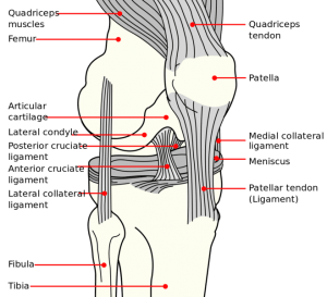
Knee anatomy
- Anterior Cruciate Ligament
- Limits anterior translation of tibia
- 75% of all hemarthroses are caused by disruption of ACL
- Posterior Cruciate Ligament
- Limits posterior translation of tibia
- Isolated injuries are rare
- Medial Collateral Ligament
- Provide restraint against valgus (outward) stress
- Lateral Collateral Ligament
- Provide restraint against varus (inward) stress
Clinical Features
- Acute trauma and pain to knee
Differential Diagnosis
Acute knee injury
- Knee dislocation
- Knee fractures
- Meniscus and ligament knee injuries
- Patella dislocation
- Patellar tendonitis
- Patellar tendon rupture
- Quadriceps tendon rupture
Nontraumatic/Subacute
- Arthritis
- Gout and Pseudogout
- Osgood-Schlatter disease
- Patellofemoral syndrome (Runner's Knee)
- Patellar tendonitis (Jumper's knee)
- Pes anserine bursitis
- Popliteal cyst (Bakers cyst)
- Prepatellar bursitis (nonseptic)
- Septic bursitis
- Septic joint
- DVT
Evaluation
Ottawa knee rules
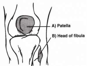
Ottawa knee rules points of tenderness
X-ray is only required in patients who have an acute injury and one or more of the following:
- Age >55
- Isolated tenderness of the patella
- Tenderness at the fibular head
- Inability flex to 90 degrees
- Inability to walk 4 steps BOTH immediately after the injury and in the ED
Management
- If xrays are positive (when indicated)
- Treat underlying condition
- If xrays are negative or not indicated Ottawa knee rules
- Do full knee exam to check for ligamentous/meniscal instability:
- Negative exam → RICE
- Positive exam or unable to evaluate secondary to pain/swelling → knee brace + RICE
- Do full knee exam to check for ligamentous/meniscal instability:
Disposition
- Depends on diagnosis; most often results in outpatient ortho referral
See Also
External Links
References
This article is issued from
Wikem.
The text is licensed under Creative
Commons - Attribution - Sharealike.
Additional terms may apply for the media files.