Acetabular pelvic fractures
Background
- Fractures usually occur when head of femur forced into acetabulum
- Obvious when displaced, subtle non-displaced
Anatomy
- Anterior column-anterior acetabulum to pubic ramus
- Posterior column- posterior acetabulum to ischial ramus
- Anterior and posterior columns merge to form acetabular dome= weight bearing portion
- Fractures involving acetabular dome require operative fixation
Clinical Features
- Pelvic pain after trauma (low energy for elderly, high energy for young)
Differential Diagnosis
Evaluation
Radiographically
- Consider obtaining AP, Judet, and inlet/outlet films
- Iliopubic line extends from ilium to superior pubic ramus
- Ilioischial line- extends from ilium to ischial ramus forming radiographic teardrop, "U" shaped, on AP pelvis
- Quadrilateral plate forms medial wall of acetabulum

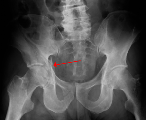
Right acetabular fracture (arrow)
Fractures Types
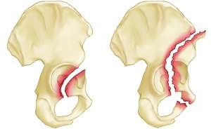
(Left) Anterior wall fracture, (right) anterior column fracture
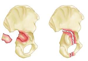
(Left) Posterior wall fracture, (right) posterior column fracture
(Left) Posterior wall transverse fracture, (right) T-shaped fracture
- Anterior column
- Posterior column
- Transverse
- T or Y-shaped
- Posterior rim
- Anterior Wall
Management
- Early ortho consultation and hospital admission is indicated for all
Disposition
- Admission
See Also
References
This article is issued from
Wikem.
The text is licensed under Creative
Commons - Attribution - Sharealike.
Additional terms may apply for the media files.